Virtual cadaver
Explore digital human anatomy through interactive dissection and structural selection. Faculty can build an explanation progressively, moving from a whole-body view to the region being discussed.
 Let’s talk ↗
Athena Table
Athena Table (model ATLV1) is an educational virtual anatomy table by Axes Medverse. The human introduction identifies Version 2.0 and the veterinary introduction identifies Version 1.0. Built on 3D Slicer and powered by Athena Hub, it brings human and veterinary anatomy together on a shared touchscreen.
Plan a demonstration ↗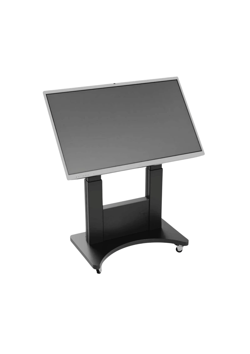 Athena Table / LEARN THROUGH EXPLORATION
Athena Table combines an interactive display and movable teaching hardware with Athena Hub’s modular anatomy environment. The table gives groups a common surface for exploring spatial relationships, comparing imaging with anatomy and discussing structures together. Instead of treating hardware and software as separate purchases, institutions can plan a configuration around their room, curriculum and teaching workflow.
A good fit for: Anatomy departments, medical and allied-health colleges, veterinary programmes and simulation centres seeking a shared digital anatomy environment.
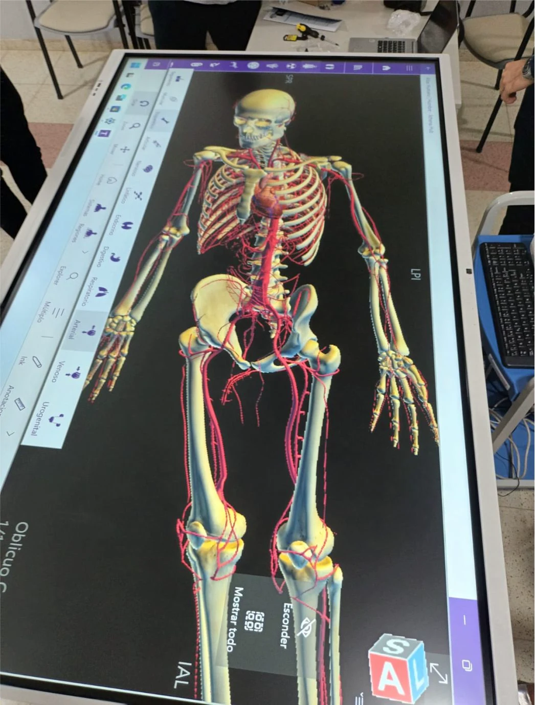
Move from a large display view to hands-on exploration around the table. A teacher can guide an anatomical explanation, invite students to identify structures and connect a selected region with the images being discussed. Plan the number of learners around each table so everyone can see and participate.
Explore digital human anatomy through interactive dissection and structural selection. Faculty can build an explanation progressively, moving from a whole-body view to the region being discussed.
Explore labelled structures in the human atlas and compare anatomy across eight animal groups: bovine, equine, porcine, feline, canine, avian, rodent and anuran.
Use DICOM views for CT, MR and ultrasound, with PACS integration and 2D multiplanar reconstruction (MPR). Confirm connectivity, permissions and data handling with your institution before using imaging datasets.
Explore volumetric, iso-surface, maximum intensity projection (MIP) and X-ray views. Compare the representations to discuss how each view reveals different features of a teaching dataset.
Create reports and print views alongside annotations and measurements. Educators can use these tools to document observations from a teaching exercise.
The veterinary atlas covers bovine, equine, porcine, feline, canine, avian, rodent and anuran anatomy. Use side-by-side teaching activities to compare structures across animal groups.
On-screen annotation and measurement tools help an educator highlight a relationship, record an observation or guide a classroom discussion. These tools support teaching; the table is presented here for educational use.
The supplied Version 1.0 brochure lists a motorised stand with remote control, a computer and 360-degree lockable wheels. Display size, room access, viewing positions, power and installation requirements should be agreed before ordering.
Athena Hub brings atlas content, virtual cadaver and medical images into a modular environment. Its mosaic view supports looking at different representations within one application. Faculty can connect a radiological slice with a structural explanation and use annotations to direct the discussion. Ask which datasets and modules are supplied with your selected edition.
Select the anatomical region, learning outcome and image material before the session. Confirm that the chosen content is available in your licensed configuration.
Rotate, scale and isolate structures; use annotations and image views to guide a focused explanation. Assign learners a question rather than asking them only to watch.
Relate the visual exploration to classroom material, clinical context or veterinary anatomy. Check understanding through explanation, comparison or identification tasks.
Share your curriculum, learner numbers, room layout, intended teaching activities and existing equipment. We can then discuss the appropriate solution and the details that need to be confirmed.
| Display & movement | 55–86 inch display options, multi-touch technology, a motorised stand with remote control, an integrated computer and lockable wheels are listed in the brochure. |
| Anatomical study | Labelled animal atlas content across eight animal groups, plus a human virtual cadaver, human atlas and radiology. |
| Interactive tools | Rotate and scale anatomical views. Explore 2D MPR and 3D volumetric, iso-surface, MIP and X-ray views. Use annotations, measurements, reporting and printing to document teaching observations. |
| Content pathways | DICOM viewing (CT, MR and ultrasound) and PACS integration are listed in the brochure. Confirm the intended data and connectivity setup during planning. |
Source: supplied Athena Table Version 1.0 brochure, product code ATLV1. Final dimensions, computer specification, software licence, included modules and installation requirements are confirmed with the institution.
Model ATLV1. The human configuration includes male and female 3D anatomy, embryology, more than 3,000 labelled structures, an immersive dental atlas, tagged dissection, high-resolution views, structure search, measurements and reconstruction.
Model ATLV1. Explore eight animal groups, with DICOM, MPR and 3D imaging workflows described in the veterinary introduction. Check species and datasets for your teaching programme.
Combine virtual dissection, a human atlas and cross-sectional views in one lesson. Isolate a region, compare structures and annotate the view during group teaching.
Teach the relationship between sectional imaging and 3D anatomy through DICOM viewing and reconstructions. Ask about case libraries, modalities and the workflow for approved teaching datasets.
Connect rendered volumetric views with available cytology and slide content. Use these modes to move between tissue and gross anatomy in the same teaching sequence.
Use veterinary content for comparative lessons and prepare approved institution-specific material. Confirm the modules, licence, hardware and import capabilities when specifying your table.
Athena Table is built on 3D Slicer (© Brigham and Women’s Hospital) for educational use. Athena Hub, the software powering the table, is owned by Medical Harbour, Brazil.
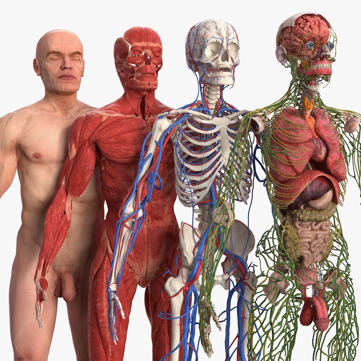
Explore structures layer by layer on the table. Faculty can guide a group through a shared view and compare labelled anatomy as the lesson progresses.
Use the mosaic workspace to compare atlas, virtual cadaver and DICOM views on Athena Table. Move between imaging slices, multiplanar views and 3D reconstructions. Use measurements and annotations to connect the imaging and anatomy lesson.
Select human or veterinary teaching material for the curriculum. Human content includes embryology and dental anatomy. The veterinary configuration covers eight animal groups.
Prepare a teaching sequence around structure recognition, relationships and group discussion. Confirm the included datasets and content modules for your institution.
Athena Table is a trademark used by Axes Medverse Private Limited. Athena Table is built on 3D Slicer (© Brigham and Women’s Hospital) and intended strictly for educational use. Athena Hub software is owned by Medical Harbour, Brazil. The product may be classified under HS Code 9023, subject to applicable customs classification rules. Confirm included modules and final specifications in writing.